Upper Leg Tendon Anatomy - One More Plate: How the Hamstrings Work / Anatomy of leg muscles and tendons muscle anatomy upper leg.
byAdmin-
0
Upper Leg Tendon Anatomy - One More Plate: How the Hamstrings Work / Anatomy of leg muscles and tendons muscle anatomy upper leg.. Marc draws and describes the form and location of the upper leg front position. What are the functions of patella. Understanding the function and anatomy of the peroneus longus can help you make the best choices for your care if you have suffered and injury there. • flatfoot deformity • flexible hindfoot • normal forefoot. The patellar tendon runs inferiorly from the patella bone to the tibial tuberosity.
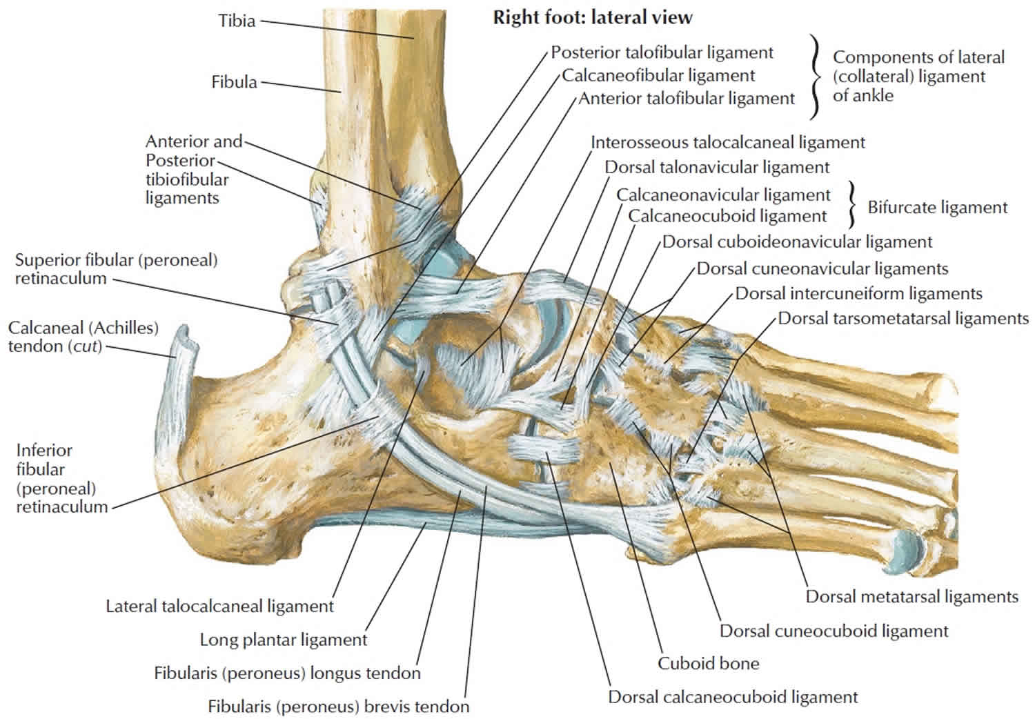
It is located from below the knee to the heel and helps in stabilizing the. • flatfoot deformity • flexible hindfoot • normal forefoot. Muscle/tendon inflammation and pain along anterio… An anatomical and biomechanical study. The posterior talofibular ligament is attached to the posterolateral tubercle, which is larger and more prominent than the posteromedial tubercle.
Upper limb anatomy 4 from www.edoctoronline.com The patella is a large sesamoid (a bone within a tendon) bone the medial and lateral parts of quadriceps femoris descend on either side of the patella and are inserted onto the upper anterior surface of the tibia. There is no real division between the core and the upper leg; Spicermanyt at checkout for 40% off this tutorial! Muscle/tendon inflammation and pain along anterio… A collection of anatomy notes covering the key anatomy concepts that medical students need to learn. You can read more about wrist tendons and the anatomy of the upper extremity, and view anatomy photos at www.handcare.org. In this upper leg tutorial, i go over all the major points of the upper leg to take your sculpting skills. Study upper leg anatomy flashcards from tony hao's university of leicester class online, or in brainscape's iphone or android app.
Blood supply to the foot.
Hands are outstretched, holding onto the handles of the bench. Muscle/tendon inflammation and pain along anterio… By spicer mcleroy in tutorials. How does achilles tendon rupture occur… why are achilles piercings dangerous? The pads of the machine are situated at the achilles tendon. Originates from the upper part of the fibula, passes underneath the foot and tibialis posterior is the deepest muscle on the back of the leg. Tendons are thick bands of tissue that connect muscles to bone. Mnemonics that can be used to remember the anatomy of the ankle tendons from anterior to posterior as they pass posteriorly to the medial malleolus of the tibia under the flexor retinaculum in the tarsal tunnel include: Spicermanyt at checkout for 40% off this tutorial! The artist's guide to the.,muscles that lift the arches of the feet and more. Fascia of the upper limb. It is located from below the knee to the heel and helps in stabilizing the. Concept 3d illustration back upper leg human anatomy.
Palmar region , arteries (illustrations: Related posts of muscle anatomy upper leg. Tendons are thick bands of tissue that connect muscles to bone. Lateral (fibular) collateral ligament (fcl) upper part middle part lower part popliteus tendon (pt) upper part i. Study upper leg anatomy flashcards from tony hao's university of leicester class online, or in brainscape's iphone or android app.
Calcaneus bone anatomy, function, calcaneus pain ... from healthjade.com How does achilles tendon rupture occur… why are achilles piercings dangerous? ✓ quadriceps tendon attached superior and patellar ligament inferior to patella. The tendons that control movement in your hands, wrists and fingers run through your forearm. What are the functions of patella. Muscle/tendon inflammation and pain along anterio… Spicermanyt at checkout for 40% off this tutorial! Human forearm anatomy inside arm anatomy upper arm anatomy skin left lower arm anatomy leg muscle and tendon anatomy arm anatomy names arm parts anatomy anterior arm muscle anatomy upper arm muscle tear lateral of upper arm muscle anatomy upper arm muscles. Fascia of the upper limb.
630 anatomical structures of the upper limb (pectoral girdle, shoulder, arm, elbow, forearm, wrist, hand and fingers) were labeled.
This mri wrist coronal cross sectional anatomy tool is absolutely free to use. They are innervated by the tibial nerve, a terminal branch of the sciatic nerve. Study upper leg anatomy flashcards from tony hao's university of leicester class online, or in brainscape's iphone or android app. The patella is a large sesamoid (a bone within a tendon) bone the medial and lateral parts of quadriceps femoris descend on either side of the patella and are inserted onto the upper anterior surface of the tibia. Tendons are thick bands of tissue that connect muscles to bone. Alas, anatomical name changes occur slowly over time and the traditional peroneus name continues to be used. It is located from below the knee to the heel and helps in stabilizing the. Superficial veins of upper limb , anatomy : A collection of anatomy notes covering the key anatomy concepts that medical students need to learn. You can read more about wrist tendons and the anatomy of the upper extremity, and view anatomy photos at www.handcare.org. Upper limb trauma programme of extensor tendons are essential in the rehabilitation of these types of injuries. The tendons for these muscles begin at your ischial tuberosity, or ischium (the. • flatfoot deformity • flexible hindfoot • normal forefoot.
Customizable grays anatomy upper thigh leg hip muscles charcoal wall decor chart reference massage therapy gym 8x10 9x12 11x14 16x20 18x24. What are the functions of patella. Use the mouse scroll wheel to move the images up and down alternatively use the tiny arrows (>>) on both side of the image to move the images. Lie prone on a hamstring curl machine. The artist's guide to the.,muscles that lift the arches of the feet and more.
Muscles in the Lateral Compartment of the Leg - TeachMeAnatomy from teachmeanatomy.info Originates from the upper part of the fibula, passes underneath the foot and tibialis posterior is the deepest muscle on the back of the leg. Spicermanyt at checkout for 40% off this tutorial! Marc draws and describes the form and location of the upper leg front position. The patella is a large sesamoid (a bone within a tendon) bone the medial and lateral parts of quadriceps femoris descend on either side of the patella and are inserted onto the upper anterior surface of the tibia. The pads of the machine are situated at the achilles tendon. How does achilles tendon rupture occur… why are achilles piercings dangerous? Collectively, the muscles in this area plantarflex and invert the foot. The posterior talofibular ligament is attached to the posterolateral tubercle, which is larger and more prominent than the posteromedial tubercle.
Related online courses on physioplus.
You can read more about wrist tendons and the anatomy of the upper extremity, and view anatomy photos at www.handcare.org. It serves to attach the plantaris, gastrocnemius (calf) and soleus muscles to the calcaneus (heel) bone. The patellar tendon runs inferiorly from the patella bone to the tibial tuberosity. Related online courses on physioplus. 3d illustration back fit strong human anatomy. Muscle/tendon inflammation and pain along anterio… Percutaneous achilles tendon lengthening is performed in the operating. An anatomical and biomechanical study. Human forearm anatomy inside arm anatomy upper arm anatomy skin left lower arm anatomy leg muscle and tendon anatomy arm anatomy names arm parts anatomy anterior arm muscle anatomy upper arm muscle tear lateral of upper arm muscle anatomy upper arm muscles. Upper leg muscles common names archives anatomy body. When a muscle contracts, the tendon pulls on the bone causing the joint to move. Mnemonics that can be used to remember the anatomy of the ankle tendons from anterior to posterior as they pass posteriorly to the medial malleolus of the tibia under the flexor retinaculum in the tarsal tunnel include: Palmar region , arteries (illustrations: